Radiography and Periodontal Diagnosis
Radiographs are often used to supplement diagnostic information gathered through examination of the soft tissues.
The most commonly used radiographs in general dental practice are bitewings and periapicals;however, an increasing number of practices are also taking OPG's in their own surgeries.
Types of radiographs
-
Bitewing radiographs
Bitewing radiographs are taken to show the proximal surfaces of the teeth and the crest of the alveolar bone of both the maxilla and the mandible on the same film. While they are used primarily to detect interproximal decay, they can also provide some information on the patient's periodontal status. The height of the interproximal alveolar bone margin relative to the cemento-enamel junction can be observed. Also, deposits of subgingival calculus may be detected. However, the value of bitewing radiographs in the diagnosis of periodontal diseases is limited by the fact that only the coronal sections of the roots of the teeth are observed, and they are limited to the molar-premolar regions.
In younger individuals, careful observation of the alveolar bone height around first permanent molars may help detect those individuals at risk of early-onset forms of periodontitis (juvenile periodontitis and rapidly progressive periodontitis). However, radiographs should be used only as a supplement to a careful clinical examination using a periodontal probe around such sites, as up to 30 per cent of bone may be lost before it is evident radiographically.
-
Periapical radiographs
Periapical radiographs are frequently used not only to aid the differential diagnoses of patients' presenting symptoms, but also to screen for otherwise undetected pathological processes of the teeth and surrounding alveolar bone.
In the diagnosis of periodontal diseases, periapical radiographs can provide useful information that cannot be obtained through examination of the soft tissues alone. Such information includes:
Teeth
- Clinical crown-root ratio: in essence, this term refers to the ratio between the length of the root that is surrounded by bone and the remainder of the tooth.
- Shape and size of the crown and root: a tooth with a small crown and a long root has a better prognosis than one with a large crown and a short root. A tapered root has less surface area for periodontal attachment than a blunt root.
- Position of the roots in multirooted teeth: in multirooted teeth, roots close together present a poorer prognosis than those which are widely separated.
- Position of a tooth in relation to adjacent teeth: open contact points or close proximity of adjacent teeth can be seen in radiographs, and may highlight areas where periodontal problems are occurring.
- Presence of callculus: both subgingival and supragingival calculus deposits can be seen on periapical radiographs.
- Presence of root resorption: internal or external root resorption can be detected.
- The contours and margins of restorations: the relationship between interproximal overhangs and/or poorly contoured restorations, and loss of periodontal bone can be screened by radiographic examination.
- Fractures of the root: a tooth with a horizontal or vertical root fracture can present with periodontal symptoms.
- Foreign bodies and root tips: these may produce or aggravate periodontal lesions and can best be detected in radiographs.
- Pulpal anatomy and pathology: the shape of the pulpal chamber and root canals can be seen, as well as pulpal pathology.
Bone
The pattern of bone loss around individual teeth can be determined only through examination of radiographs. Periapical radiographs, using the long cone paralleling technique, provide the most accurate representation of the height of the bone in relation to the CEJ, and to the actual length of the tooth. In the examination of bone features in periapical radiographs as part of periodontal diagnosis, special attention is paid to:
- Pattern of bone loss: is the bone loss horizontal or vertical?
- Extent of bone loss: is the bone loss generalised over the dentition or localised to certain teeth?
- Severity of bone loss: this can be expressed in percentage terms, taking normal bone height to be just below the CEJ, and accounting for the length of the root.
- Furcation involvement: is there evidence of radiolucencv in furcation areas?
- Lamina durn: the significance of the lamina dura is unclear. Whereas its presence indicates good supporting bone, its absence does not always signify pathology.
- Periodontal ligament space: a widening of the periodontal ligament space may indicate that the tooth is subject to occlusal forces or is mobile. It may also be an early sign of pulpal inflammation, therefore a careful clinical examination is necessary to make a diagnosis.
-
Panoramic radiographs
Panoramic radiographs provide a general view of the oral structures, and are useful for screening bone loss patterns in general. They are not suitable for accurate assessment of the degree of bone loss associated with individual teeth, as there is severe distortion and the outline of the bone margin is often unclear due to superimposition of intervening structures.
Using radiographs
-
Technical problems with radiographs
Variations in x-ray technique that produce distortion, coupled with the projection of a three-dimensional object onto a two-dimensional plane, limits the diagnostic value of the radiograph. The bone level, the pattern of bone destruction, the radiodensity and trabecular pattern of the interdental bone, all are modified by altering the exposure and development time, the type of film and the x-ray angulation. These features must be considered when trying to compare pre- and post-treatment radiographs. The long cone paralleling technique is recommended for all periapical radiographs taken for periodontal diagnostic reasons, as it produces the most realistic image of the alveolar bone.
The bisection of the angle technique increases the angle of projection and makes the bone margin appear closer to the crown; the level of the facial bone is distorted more than the lingual. Shifting the cone mesially or distally without disturbing the horizontal plane pr-qjects the x-rays obliquely and changes the image of the interdental bone, and may distort the extent of furcation involvement. For these reasons, it is strongly recommended that you use beam aligning holders for taking long-cone parallel technique radiographs. (for example the RINN aligning devices, available through Dentsply). This technique also allows for more standardised comparisons of radiographs over time even if taken by different operators.
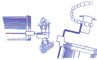
Rinn device - posterior area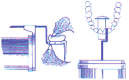
Rinn device - anterior area
-
Recording of radiographic findings
It is important to note the findings of your radiographic examination in the patient's treatment record, and to mount and store the radiographs for future reference. Notations in the treatment record should indicate:
- the date the radiographs were taken
- the type of radiographs taken
- the reason for taking the radiographs
- diagnostic information gained from examination of radiographs
- further diagnostic tests that may need to follow up any pertinent radiographic findings.
This material has been compiled with the assistance of Dr Louise Brown, Lecturer in Periodontics at the University of Melbourne.