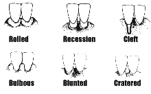
Gingival Recession
The concept of becoming "long in the tooth" appears to be accepted by many patients as an inevitable part of the aging process.
Indeed, many adult patients will have areas of gingival recession that are stable and require no specific intervention. However, in some patients, recession may lead to the exposure of an unacceptable amount of root surface, causing aesthetic problems, dentinal hypersensitivity, potential root canes, as well as mucogingival problems.
The attached gingiva is defined as the portion of the gingivae extending from the base of the gingival crevice to the mucogingival line. It is firm, dense, sometimes stippled, and tightly bound down to the underlying periosteum, tooth and bone. The width of the attached gingiva was studied by Bowers (1963), who concluded:
- the width of the attached gingiva varies with each tooth
- there is great variation in the width of attached gingiva between patients, but the pattern of variation is consistent, varying between 1-9 mm
- there is an increase in the mean width of the attached gingiva from the deciduous dentition to the adult dentition
- the mean widths of attached gingiva for individual teeth are approximately the same for males and females
- there is a greater overall width of attached gingiva in the maxilla than the mandible
- teeth that are prominent in the arch such as the lower cuspids have a narrower zone of attached gingiva
- teeth rotated labially have a narrower zone than the corresponding tooth on the opposite side that is well aligned
- high frena and muscle attachments are usually associated with narrow bands of attached gingiva
- it is possible to maintain clinically healthy gingiva despite a very' narrow zone of attachment (less than 1 mm).
-
Causes of recession
Before recession can occur, a wounding of the band of attached gingiva must. occur. The actual process by which it occurs remains questionable. Baker and Seymour (1976) described the process in rats where minor wounding or repeated inflammation caused by plaque resulted in downgrowth in rete pegs from the facial surface of tile gingiva eventually meeting epithelial strands which grow into the inflamed tissue from sulcular epithelium on the sulcus side. The union of the two epithelia creates an area of weakness that splits open when further wounding occurs.
Predisposing factors
- Inadequate attached gingiva
- high frenum attachment
- shallow vestibule
- Malpositioning of teeth (prominent roots)
- Osseous dehiscence
Precipitating factors
- Vigorous brushing, especially with suffer brushes
- Laceration
- Recurrent inflammation
- latrogenic factors
- Inadequate attached gingiva
-
'Inadequate' attached gingiva
An adequate band of attached gingiva is that amount that is sufficient to prevent, recession. An inadequate hand is one that will not prevent recession. Several studies have been undertaken to find a magic figure by which adequacy can be gauged, with some authors concluding that a band of at least I mm is required to maintain gingival health, while others have found that a lack of attached gingiva need not result in an increase in recession, if oral hygiene is excellent.
A number of factors have to be considered in relation to the need for surgical intervention where there "inadequate" attached gingiva:
- the age of the patient
- which teeth are involved
- existing or potential aesthetic problems
- existence of recession with consequent aesthetic or sensitivity problems
- the oral hygiene practices of the patient
- the restorative needs of the patient.
In essence, the whole question of determining how much is an adequate amount of attached gingiva is one of clinical judgement. Dentists have a responsibility to inform patients of the likelihood of further recession if no intervention is performed, and this advice to the patient must result from a thorough examination of the tissues, a review of the patient's oral hygiene practices and the patient's future dental needs.
-
Monitoring recession
Accurate measurements of the amount of recession that a patient presents with at their initial examination can be difficult to record. A periodontal probe can be used to measure from the CEJ to the gingival crest at a number of sites around a tooth. However, if the tooth is restored or decayed at the CEJ, this figure may not be very reproducible. Study models are a useful way of capturing the ginvigival contours, and they have the advantage that they can be used lo educate the patient about the problems of oral hygiene in relation to recession. Clinical photography can provide an excellent, record of not only the contour of the recession, but also the degree of inflammation of the tissues. Changes to the level of recession can then he determined against the baseline measurements.
-
Treatment
As with most aspects of dentistry, prevention should be our primary focus. Instructing patients to use a non-traumatic method of tooth brushing that maintains the gingival tissues in health is essential from a young age. Care must be exercised in placing restorations near the gingival margins so that the tissues are not lacerated during cavity preparation, and that the restoration is neither under- or overcontoured at. the gingival margin. Orthodontic movement of anterior teeth into labial positions can be followed by gingival recession and loss of attachment, particularly if there is a plaque induced inflammation of the gingival tissues, and the gingival tissues are thin. In some patients, a gingival graft placed over susceptible sites prior to orthodontic treatment may be used to prevent recession during and after treatment, although this is still a controversial treatment decision.
Both surgical and non-surgical periodontal treatment can result in marked recession, particularly if there is loss of significant bony support, in the presence of heavily inflamed, oedematous tissues. Patients need to be warned that recession may occur as the gingival tissues heal following treatment. The decision to proceed with periodontal surgical procedures in the anterior regions of the mouth must take into account the likelihood of aesthetic and possibly phonetic problems if recession leads to loss of the interdental papillae.
A number of mucogingival surgical procedures are available to increase the width of attached gingiva. These include the use of free gingival grafts, pedicle grafts (eg. laterally re-positioned grafts, coronally repositioned flaps) and subepithelial connective tissue grafts. The use of these procedures can lead to the formation of a stable, non-inflamed band of attached gingival tissue.
-
Cited references
Bowers GM. (1963) A study of the width of attached gingiva. JPeriodontol 34: 201-209.
Baker DL, Seymour GJ (1976) The possible pathogenesis of gingival recession. A histological study of induced recession in the rat. J Clin Periodontol 3: 208-219.
This material has been compiled with the assistance of Dr Louise Brown, Lecturer in Periodontics at the University of Melbourne.

