Furcation Involved Teeth
The progression of periodontal attachment loss to involve the furcation areas of multi-rooted teeth adds a complex dimension to the diagnosis and treatment of a patient's periodontal condition.
It also alters the prognosis of the involved teeth, thus affecting the dentist's decisions regarding restorative options, such as the use of the tooth as an abutment for fixed restorative work.
-
Anatomy
Knowledge of the anatomy of multi-rooted teeth is essential in order to correctly identify the presence of furcation involvements.
Maxillary molar teeth
- the mesiobuccal roots are comparatively wide in the buccopalatal direction, and frequently have marked concavities
- the distobuccal roots are smaller in dimension, more rounded in outline and less frequently have distinct concavities
- the palatal roots are wider in the mesiodistal than in the buccopalatal direction
Maxillary premolar teeth
- most maxillary first premolars are bifurcated at the middle third of the root trunk
- the canine fossa creates a deep concavity on the mesial root that merges with the opening of the furcation
Mandibular molar teeth
- generally have two roots of similar size and length
*the mesial root is usually barbell-shaped due to mesial and distal concavities - the distal root has a mesial concavity and usually has a smaller buccolingual dimension
-
Diagnosis
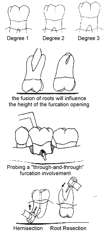
Diagnosis of furcation involvement is based upon probing and radiographic findings. Although a straight periodontal probe may be used, detection of subgingival furcations is facilitated by the use of specially designed furcation probes (Nabers No I and No 2 probes). To detect involvement, the tip of the probe is moved towards the presumed location of the furcation and then curved into the furcation area. For the mesial surfaces of maxillary molars, this is best done from a palatal direction, as the mesial furcation is located palatal to the midpoint of the mesial surface. The distal furcation of maxillary molars is located more towards the midline, and may be detected from a buccal or palatal approach. Furcation involvement may be classified into three degrees, depending upon the extension of destruction in the horizontal direction.
Degree 1: initial involvement. The furcation opening can be felt upon probing. The horizontal probing depth is less than one third of the width of the tooth.
Degree 2: partial involvement. The horizontal loss of periodontal support exceeds one third of the width of the tooth, but does not encompass the total width of the furcation area.
Degree 3: through-and-through involvement. The probe passes through the entire dimension of the furcation.
Radiographically, the presence of a furcation involvement. is evident as a radiolucent area in the vicinity of a furcation. However, due to the superimposition of roots and bony-structures, there may be little or no radiographic evidence of Degree I and 2 furcation involvements.
-
Differential diagnoses
The progression of periodontitis to involve the furcation area is not the only cause of bone loss within a furcation area. The lesion may be endodontic in origin, or may arise following perforation of the furcation during endodontic treatment or post preparation. Occasionally, a root fracture will involve the furcation area. A thorough examination of the tooth is required to form a diagnosis of the problem.
-
Treatment options
There are a number of options available to manage teeth with furcation involvements. Whatever the choice of treatment, it is necessary to ensure that healing results in a morphology in the furcation area that is optimal for oral hygiene to be performed effectively. In most teeth with Degree I furcation involvement, this can be achieved by thorough scaling and root-planing. A problem which arises in root-planing the furcation area is that the entrance of the furcation is often narrower than the width of the curette blade. The use of older instruments that have been Finely sharpened to a narrow blade may be useful, or else there are ranges of curettes with very narrow heads (eg the Gracey Minis or 'after-five' series of curettes). The use of fine ultrasonic cleaner tips, for example the Cavitron EWP-IO, EWP-12R and EWP-12L series allows for access into narrow furcations.
Surgical access is often required to adequately debride teeth with Degree 2 or 3 furcation involvements. At the time of surgery, odontoplasty may be performed in order to widen the furcation opening to the size of an interdental brush. This may also involve some minor osteoplasty to recontour the bony margins of the furcation area. The flap may be apically repositioned in order to allow access for cleaning by the patient. Extensive recontouring of the roots must be avoided, however, as this can lead to problems of hypersensitivity, may adversely affect pulp vitality, and it may increase the risk of root caries. These problems may be minimised by the use of daily fluoride mouthwashes, and the topical application of fluoride varnishes or desensitising agents.
The development of guided tissue regeneration techniques has provided an important expansion to the range of treatment options for teeth with furcation involvement. This procedure has been most successful in the management, of mandibular molars with Degree 2 furcation involvements, with more limited success in maxillary molar teeth.
If teeth with deep Degree 2 or 3 furcation involvement fail to respond to the above range of therapies, then root resection is often the next treatment of choice. It is preferable that the endodontic treatment of the root to be retained following root resection or tooth hemisection is completed prior to surgery. If no root can be preserved, then extraction may be the only option. Extraction may also be performed when the maintenance of the affected tooth is not essential for a functioning dentition.
-
Prognosis
The detection of a furcation involvement can affect the prognosis of the tooth. Therefore, as part of the initial assessment of every patient, it is essential to judge the location of the furcations and to prevent them from becoming exposed as a result of treatment or from disease progression. Studies examining the long term prognosis of teeth with furcation involvements have shown that maxillary second molars are most commonly lost, followed by maxillary first premolars. However, these studies also demonstrate that the vast majority of teeth with furcation involvements can be successfully treated and maintained over many years.
This material has been compiled with the assistance of Dr Louise Brown, Lecturer in Periodontics at the University of Melbourne.

